20 Jun 2026
Foot Drop: Causes from Nerve, Spine and Brain and How Amritsar Neurologists Evaluate

22 May 2025
Call +91 80788 80788 to request an appointment.
Cirrhosis is a progressive liver disease characterized by irreversible liver damage due to various underlying causes, including chronic alcohol abuse, viral hepatitis, and metabolic disorders. As the liver becomes increasingly scarred, its ability to function deteriorates, leading to severe health complications. Recognizing cirrhosis early is crucial for effective management and improving patient outcomes. Advanced imaging techniques play a pivotal role in the diagnosis, assessment, and monitoring of cirrhosis. This article aims to explore these techniques and their importance in cirrhosis assessment, especially here in Punjab.
Cirrhosis can be caused by a variety of factors, each contributing to liver cell injury and subsequent fibrosis. Common causes include:
Patients with cirrhosis may exhibit a range of symptoms, including fatigue, weakness, jaundice, swelling in the legs and abdomen, and unexpected weight loss. Advanced stages might lead to complications such as liver failure or hepatocellular carcinoma (liver cancer), making timely assessment and management crucial.
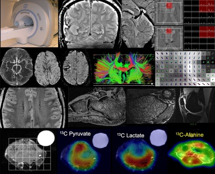
Advanced imaging techniques are essential tools for liver health assessment and cirrhosis evaluation. They not only help in diagnosing the condition but also in determining the extent of liver damage and monitoring disease progression. The following are some leading advanced imaging techniques utilized for cirrhosis assessment at Livasa Hospitals in Punjab:
Each imaging modality offers unique advantages and insights into liver health, enabling tailored management strategies for patients suffering from cirrhosis.
Ultrasound is often the first-line imaging modality used in cirrhosis assessment due to its non-invasive nature and accessibility. It employs sound waves to create images of the liver and surrounding structures. This technique is effective in:
The availability of ultrasound in Punjab makes it an essential tool for early detection and routine follow-up, particularly in high-risk individuals.
The CT scan provides a more detailed cross-sectional view of the liver than ultrasound does. This imaging technique is beneficial for:
CT scanning in conjunction with other imaging modalities helps healthcare providers develop comprehensive treatment plans, improving patient care at Livasa Hospitals.
Magnetic Resonance Imaging (MRI) stands out due to its ability to provide high-resolution images of the liver. This technique is particularly useful for:
MRI is also advantageous due to its lack of ionizing radiation, making it a preferred choice for certain populations, such as children or patients needing repeated imaging.
Liver fibrosis is a key factor in cirrhosis progression. Elastography is an advanced imaging technique that measures liver stiffness non-invasively, quantifying the degree of fibrosis. There are two main types of elastography:
Both methods have proven effective in staging liver disease and determining treatment urgency, providing critical information that guides clinical decision-making. In Punjab, availability of these technologies at Livasa Hospitals enhances patient care through precise assessments.
When selecting an imaging technique for cirrhosis assessment, it is essential to understand the unique advantages of each modality. The table below offers a comparative overview:
| Imaging Modality | Advantages | Limitations |
|---|---|---|
| Ultrasound | Non-invasive, low-cost, readily available | Operator-dependent, limited detail |
| CT Scan | High-resolution images, good for complex cases | Involves radiation exposure |
| MRI | Detailed views, no radiation | Higher cost, longer wait times |
| Elastography | Non-invasive stiffness assessment | Limited by absorptive fat, operator skill |
Despite the differences, combining these techniques enables thorough assessment and effective management of cirrhosis at Livasa Hospitals throughout Punjab.
The landscape of **advanced imaging techniques** continues to evolve, offering new possibilities for cirrhosis assessment and management. Recent advancements include:
Staying at the forefront of these innovations is essential for Livasa Hospitals, ensuring that our patients benefit from the best available liver health assessment options in Punjab.
In conclusion, advanced imaging techniques for cirrhosis assessment are indispensable for timely diagnosis and effective management. The applications of ultrasound, CT scans, MRI, elastography, and more provide comprehensive insights into the liver's condition, paving the way for improved clinical outcomes. By leveraging these technologies, Livasa Hospitals in Punjab stands committed to enhancing liver health and empowering patients to maintain optimal well-being.
If you or a loved one is at risk for liver disease, don’t hesitate to reach out to our gastroenterology specialists for a thorough evaluation. Early detection can be the key to effective management and a healthier future.
To schedule your liver health assessment or learn more about advanced imaging techniques, book your appointment at Livasa Hospitals today or consult our specialists.
Foot Drop: Causes from Nerve, Spine and Brain and How Amritsar Neurologists Evaluate
When Neck or Back Pain Needs MRI and Neurosurgeon Opinion in Amritsar
Neuropathy in Diabetes: Joint Care by Neurologist and Diabetologist in Amritsar
Livasa Healthcare Group Corporate Office,Phase-8, Industrial Area, Sector 73, Sahibzada Ajit Singh Nagar, Punjab 160071
| Mohali | +91-99888 23456 |
| Amritsar | +91-99887 49494 |
| Hoshiarpur | +91-99883 35353 |
| Nawanshahr | +91-75081 82337 |
| Khanna | +91-98888 05394 |
